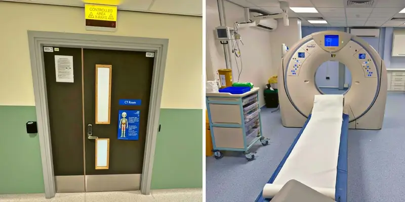
The CUH CT department is specialised in providing a range of procedures including all body CT scanning, Cardiac scans, CT Colonography, Enterography and CT Post Mortem.
We have close links with Cambridge University and support a variety of research trials with many different research institutions.
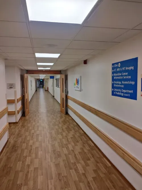
Continue along the corridor to either the CT department or PET-CT department (where we also have a CT scanner located). The above photo was taken as you leave the main Outpatient corridor; follow the corridor for about 50 metres, where you will find the door to the CT reception is on your left.
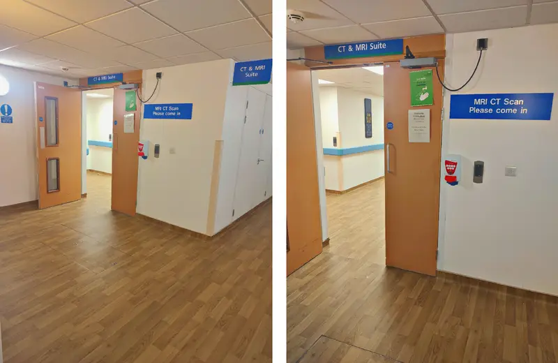
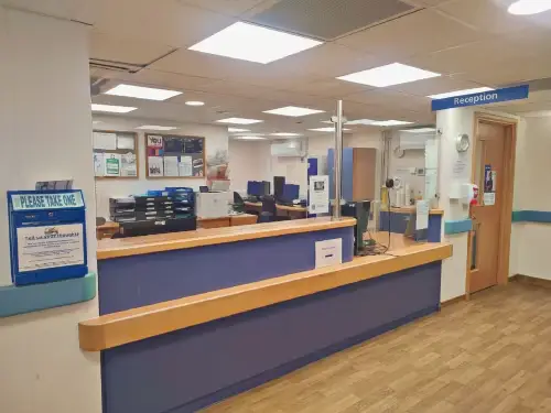
On arrival, please approach the front desk and let the administration staff know that you have arrived.
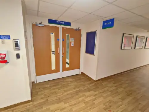
Currently we have six CT scanners at the CUH site (one located in the PET-CT department) as well as a CT units located at Sawston Medical Centre and the Ely Community Diagnostic Centre (Princess of Wales Hospital).
Please check your appointment letter carefully for further details.
Having a CT scan
A Computerised Tomography (CT) scan uses x-rays and a computer to create detailed images of the inside of your body. It is similar to how an x-ray machine works but the x-ray tube spins around you taking lots of pictures that are then re-arranged to appear as ‘slices’ through the body.
The x-rays pass through the body and hit a detector which converts the signals into pictures. Large complex computers convert all this data into slices through the body that can then be used by Radiographers and Radiologists.
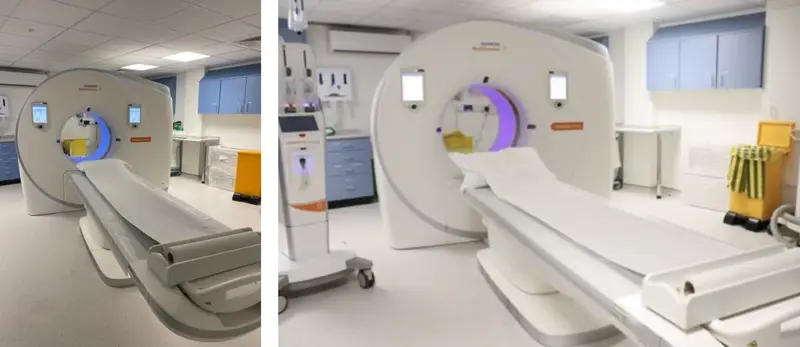
The data gathered from the scan can also be used to produce 3D images of certain body parts, for example blood vessels or a particular bone. CT scans are useful for looking at bones, lungs and other organs, and blood vessels. CT scans are also used to look at a wide range of conditions and pathologies.
The CT scanner is doughnut shaped and tends to be wider in the middle compared to an MRI scanner and not as long. You will be asked to lie on the CT table which automatically passes through the centre of the scanner, known as the gantry bore.
You may be given an injection of ‘contrast media’ or for some investigations, asked to drink a small volume of the oral ‘contrast media’ to help enhance the pictures. The contrast media allows us to see certain structures like blood vessels. Please see below for further details. The whole process of having a scan usually takes 10-15 minutes depending on the complexity of the scan being performed. The actual scan itself can take a matter of seconds though.
For patients
The Clinical Imaging Board's 'Your CT scan' poster (opens in a new tab) provides information regarding having a CT scan.
Benefits
- CT scans can give doctors information to help them diagnose a variety of conditions. The scans can help to confirm or rule out a suspected diagnosis.
- Unlike other imaging methods, a CT scan can give a detailed view of lots of different tissue types in the body, including lungs, bones, soft tissues and blood vessels.
Risks
- The CT scan involves exposure to radiation in the form of X-rays. However, the amount you are exposed to during the scan is very small. Please make your Radiographer aware of any recent CT scans you may have had, in case they make further examinations unnecessary.
- If you have contrast injected, there is a small chance that the contrast can leak outside the vein and cause temporary swelling and bruising at the site of the injection.
